|
|
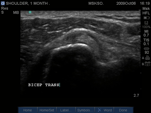
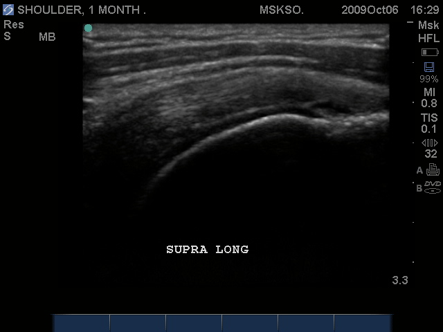
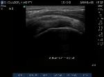
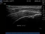
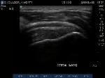
Subluxing Bicep tendon
over lesser tuberosity
|
|
|
|
 |
|
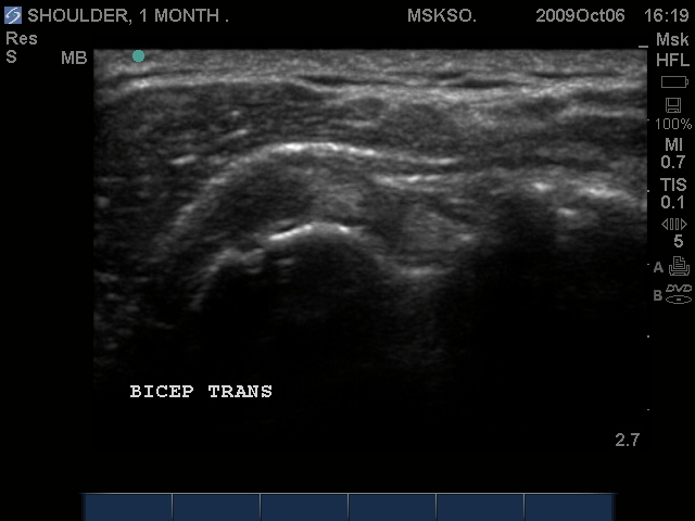
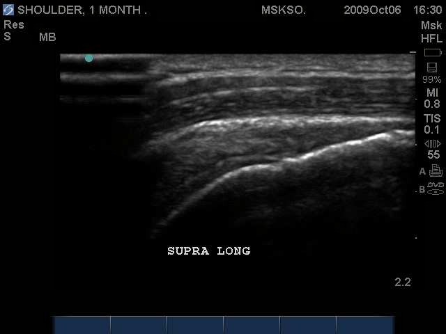
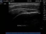
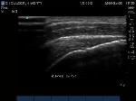
Subluxing Bicep tendon
over proximal lesser
tuberosity
|
|
|
|
 |
|
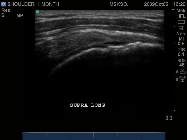
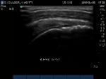
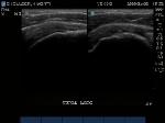
Splitting Bicep tendon
over lesser tuberosity
|
|
|
|
 |
|
|
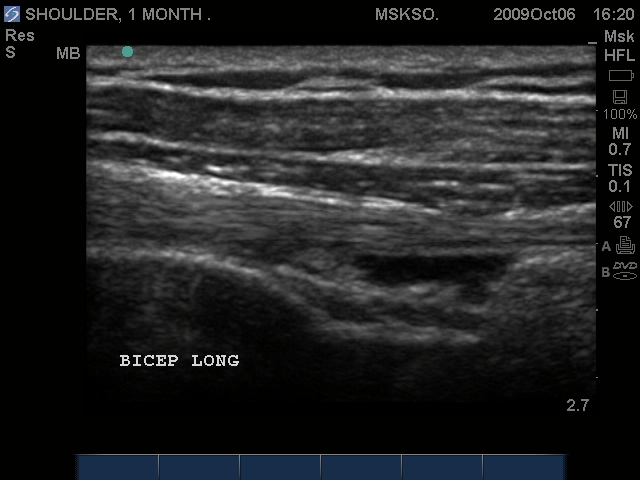
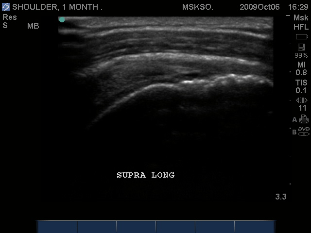
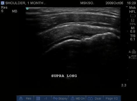
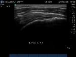
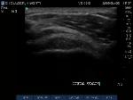
Long axis Bicep tendon
with synovial fluid at
level of surgical neck
|
|
|
|
 |
|
|
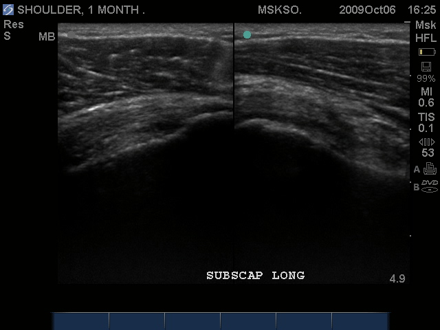
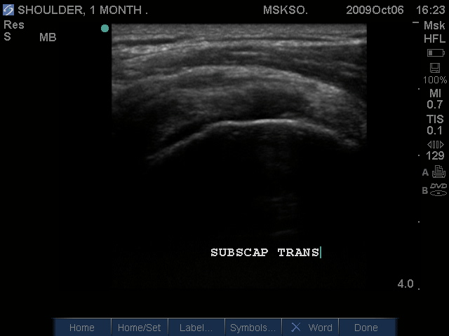
Long axis Subscapularis
tendon inferior margin;
articular surface partial tear
|
|
|
 |
|
|
Long axis (left) and
Transverse (right)
images of Subscapularis
Tear
|
|
|
|
 |
|
|
Proximal Subscapularis
tendon at tear site over
humeral head transverse
|
|
|
 |
|
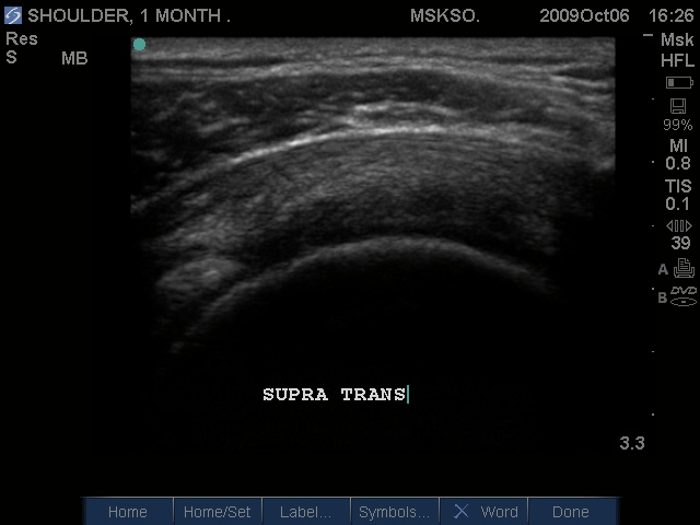
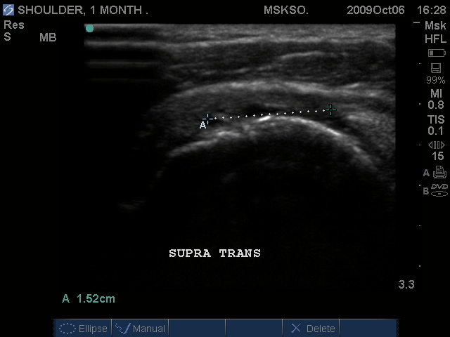
Supraspinatus tendon
transverse over humeral
head articular surface
(articular surface
changes in echotexture
to hypoechoic)
|
|
|
 |
|
"Cartilage Interface Sign"
the hyperechoic rim over
the hypoechoic articular
cartilage is not usually
seen unless a fluid layer
is between the tendon
and cartilage surface.
When the tendon is
resting normally over
cartilage the interface is
only hypoechoic.
|
|
|
 |
|
|
Supraspinatus imaged
distally over the Greater
Tuberosity. Large articular
surface tear measured
anterior to posterior,
Infraspinatus tendon is seen
at the right as a hyperechoic
tendon transverse oblique
over the posterior humeral
head.
|
|
|
|
 |
|
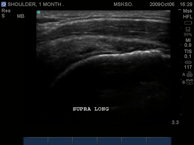
Long axis Supraspinatus
tendon over lateral
Greater Tuberosity.
(lateral greater
tuberosity is flat relative
to the anterior) Tendon
retraction is seen over
cartilage interface sign of
the humeral head.
|
|
|
|
 |
|
Slightly anterior
movement of the probe
to visualize the full detail
of the tear only for this
case study purpose.
|
|
|
|
 |
|
Anterior movement of
the probe from the prior
image shows beginning
of cortical irregularities
seen at the lateral
Greater Tuberosity
|
|
|
|
|
 |
|
Anterior Greater Tuberosity
imaged as a more
pronounced curved cortical
margin. This area of the
insertion is truly
Supraspinatus with no
Infraspinatus merger. Here
we see more cortical
irregularity deeper into the
cortex.
|
|
|
|
 |
|
|
Compression of the tear
site may reveal a more
normal cuff appearance,
in this case, the flattening
of the normally convex
appearing bursal surface
would indicate that there
is volume loss at this
level.
|
|
|
 |
|
Transverse and Long
axis images should
always be taken to
demonstrate 2 imaging
planes 90 degrees from
each other.
|
|
|
|
 |
|
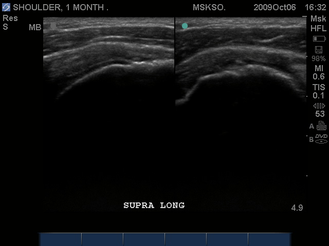
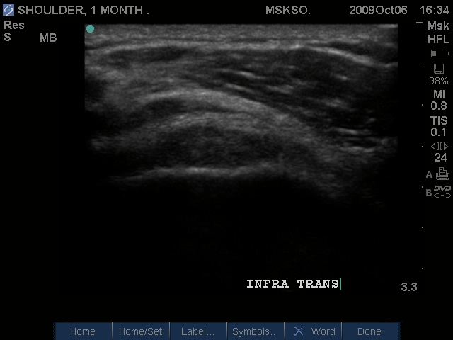
Posteriorly the
Infraspinatus tendon in
transverse plane is
intact, but shows a
bursitis that can be
traced over the greater
tuberosity. Proximal to
this site this may be a
normal myotendinous
junction appearance.
|
|
|
|
 |
|
|
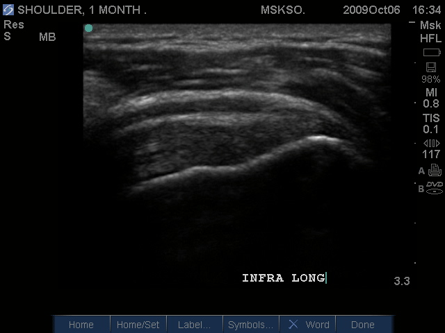
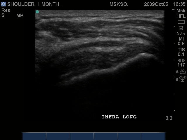
Long axis image of the
Infraspinatus tendon
over the posterior
Greater Tuberosity also
confirms bursal
enlargement.
|
|
|
|
 |
|
Compression shows this is
not simple bursal fluid.
|
|
|
|